
Clinical Motion Sensing & Biomechanical Product Fleet
Engineered for low-latency posture monitoring, quantitative functional range of motion (ROM), and seamless tele-rehabilitation streaming.
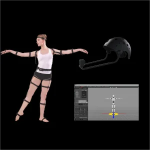
Design VDCap Full Performance Capture Entry-Level Motion Capture Kits for Beginners and Hobbyist Planning, Solution
Explore Solution →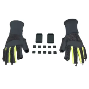
Best Inertia Motion Capture Fingers Capture Accessories Elastic Lycra Fabric Gloves for VDSuit Full(Without Sensors) Planning, Solution
Explore Solution →
Best Virtual Live Technology Empowerment Makes Online Education and Learning As Fun As Watching Cartoons Planning, Solution
Explore Solution →
Best How Do Brands Use Virtual Spokesman to Tell Good Stories: Motion Capture Technology Empowers Brand Propagation Planning, Solution
Explore Solution →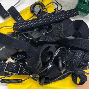
Best Full Body Motion Capture Accessories for Suit Elastic Tapes Planning, Solution
Explore Solution →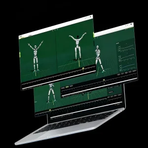
Design VDTraining: One-Stop Robot Reinforcement & Imitation Learning Platform Planning, Solution
Explore Solution →
Best Virdyn Humanoid Robot Live Event Performance Solution for Robot ce&Teleoperation Planning, Solution
Explore Solution →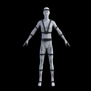
Design Virdyn VDSuit Full for Full Body Function Inertia Motion Capture Suit Planning, Solution
Explore Solution →Global Commercial & Clinical Landscape of Rehabilitation Mocap
The transition from subjective visual scoring to objective, continuous, and millimeter-level kinematic quantification is redefining global orthopedics, physical therapy, and neurological recovery protocols.
In historical physical rehabilitation frameworks, functional recovery assessments have heavily relied on ordinal, subjective metrics such as the Fugl-Meyer Assessment (FMA), the Berg Balance Scale (BBS), and qualitative goniometry. While clinically established, these observational methods present notable limitations: intra- and inter-rater variability, susceptibility to compensatory movement patterns masquerading as genuine motor recovery, and an absence of continuous kinematic telemetry outside hospital walls.
The contemporary global commercial landscape is experiencing an infrastructural pivot toward computerized motion capture systems specifically adapted for physical medicine and rehabilitation (PM&R). Driven by the confluence of aging demographics, escalating neurovascular pathologies (such as ischemic stroke and traumatic brain injuries), and the democratization of micro-electromechanical systems (MEMS), healthcare networks across North America, Europe, and Asia-Pacific are systematically embedding real-time motion capture into outpatient clinics, robotic therapy centers, and home-based decentralized care programs.
Furthermore, standard medical coding and reimbursement pathways (such as CPT codes in the United States covering computerized motion analysis, gait analysis, and remote therapeutic monitoring) are providing concrete fiscal legitimacy. Consequently, motion tracking has evolved from an academic curiosity confined to specialized biomechanics research institutes into an indispensable clinical diagnostic tool delivering measurable clinical throughput and audited patient recovery outcomes.
Strategic Clinical Shift: From Lab Bounds to Unconstrained Point-of-Care
The paradigm shift centers on moving beyond the physical boundaries of multi-camera optical gait laboratories. By leveraging medical-grade wearable inertial measurement units (IMUs) equipped with dynamic Kalman filtering and magnetic anomaly resilience, clinicians can now execute high-precision kinematic analysis in standard examination rooms, outdoor gait training surfaces, and rural community health clinics.
Technological Evolution: Optical vs. Inertial vs. Markerless CV
Understanding the clinical trade-offs across modalities enables procurement teams and medical directors to deploy the optimal tracking infrastructure for targeted pathologies.
| Evaluation Parameter | Marker-Based Optical (Stereophotogrammetry) | Wearable Inertial (IMU Sensor Fusion) | Markerless Computer Vision (Edge AI RGB-D) |
|---|---|---|---|
| Spatial Measurement Domain | Constrained calibrated volume (typically 6x6m to 10x10m). | Completely unconstrained; indoor, outdoor, ambulatory. | Field-of-view of camera array (usually 3x4m cone). |
| Angular Kinematic Accuracy | Sub-millimeter spatial; < 0.5° angular resolution. | < 0.8° - 1.2° angular tracking error with active fusion. | 1.5° - 3.5°; sensitive to clothing fit and edge occlusion. |
| Setup Latency & Donning Time | 20–45 min per patient (skin palpation, retroreflective markers). | 3–6 min (elastic biomechanical harnessing or straps). | Instantaneous zero-donning setup (ambient capture). |
| Occlusion Vulnerability | High (crutches, wheelchairs, orthoses obstruct line of sight). | Zero occlusion impact (body-worn internal sensor nodes). | Moderate to High during wheelchair or walker interaction. |
| Cost Profile & Footprint | $80,000 - $250,000; dedicated clinical facility required. | $4,000 - $18,000; portable case, accessible anywhere. | $10,000 - $40,000; standard workstations & high-speed cameras. |
| Optimal Clinical Application | Complex pediatric cerebral palsy pre-surgical planning. | Ambulatory stroke rehab, telerehab, spinal kinematics. | General geriatric triage, non-contact range of motion triage. |
Phase 1: Sensor Miniaturization & Biomechanical Constraints
Integration of low-power 9-axis tri-axial accelerometers, gyroscopes, and magnetometers within ultra-lightweight housings. Elimination of soft-tissue artifact through mathematical anatomical constraints and dynamic joint center estimation algorithms.
Phase 2: Real-Time Digital Twin & Musculoskeletal Modeling
Real-time inverse kinematics (IK) and inverse dynamics running directly on edge computing units. The raw quaternion stream drives customized OpenSim biomechanical musculoskeletal models to output tendon strain, joint contact forces, and metabolic energy consumption.
Phase 3: Closed-Loop Neuro-Modulation & Robotic Actuation
Streaming kinematic telemetry acts as the sensory input for adaptive lower-limb exoskeletons and Functional Electrical Stimulation (FES). If gait phase deviations (e.g., foot drop) exceed normative thresholds, stimulation is triggered in <15ms to restore physiological cadence.
Targeted Clinical Protocols & Localized Rehabilitation Scenarios
Engineered to address high-incidence clinical conditions across acute hospital wards, specialized rehabilitation centers, and community tele-health networks.
Neurological & Post-Stroke Rehabilitation
Stroke survivors frequently present with hemiparetic gait, circumduction, and compensatory trunk lean. Body-worn inertial sensors track spatiotemporal parameters—such as step length symmetry, stance-to-swing phase ratios, and pelvis obliquity—isolating genuine cortical recovery from motor compensation.
Orthopedic Post-Operative Kinematic Tracking
Following Total Knee Arthroplasty (TKA) or Anterior Cruciate Ligament Reconstruction (ACLR), recovery benchmarks demand precise sagittal plane knee flexion and extension dynamics. Mocap captures dynamic valgus collapse during kinetic chain squats, preventing graft failure.
Geriatric Fall Risk Stratification & Balance
Utilizing high-frequency trunk and pelvic acceleration variance to compute Center of Mass (CoM) trajectory excursions during standard Timed Up and Go (TUG) assessments. Quantitative micro-hesitations in gait cadence identify fall risks months before clinical incidents occur.
Decentralized Telerehabilitation & Biofeedback
Eliminating geographical barriers by deploying lightweight mocap garments paired with mobile computing. Patients execute guided therapeutic exercises while the interface renders real-time visual-auditory error feedback, concurrently syncing kinematic datasets to clinicians.
Macro Clinical Solutions Architecture: From Sensor to EHR
A resilient medical-grade deployment requires tight convergence across hardware ergonomics, real-time wireless telemetry, biomechanical processing engines, and clinical reporting workflows.
Modern clinical rehabilitation demands that motion capture operates not in isolation, but as a deeply embedded diagnostic layer within hospital information networks. When designing a clinical-grade motion tracking suite, the following tiered architecture ensures regulatory compliance, data security, and physician usability:
Tier 1: Ergonomic Wearable Sensor Topography
Medical-grade textile integration utilizing hypoallergenic, breathable elastic bands with silicone backing to eliminate sensor slippage during dynamic motion. Sensor pods incorporate industrial MEMS architectures calibrated across broad temperature curves, shielded against environmental electromagnetic noise from physical therapy tables.
Tier 2: Algorithmic Fusion & Skeletal Solving
Raw 6-axis / 9-axis sensor streams are packaged via 2.4GHz proprietary ultra-low latency protocols or Bluetooth Low Energy 5.3. On the local gateway, algorithms calculate orientation matrices, execute drift correction utilizing static stance phases (Zero Velocity Updates - ZUPT), and solve the biomechanical hierarchical kinematic chain.
Tier 3: HL7/FHIR Integration & Automated Reporting
Kinematic metrics are synthesized into automated clinical summary documents highlighting range of motion, symmetry index, cadence, and deviations from age-matched normative benchmarks. Secure integration with Electronic Health Records (Epic, Cerner) via standard HL7/FHIR protocols guarantees audited continuity of care.
Regulatory Compliance & Validation Pathways (FDA 510(k) & CE MDR Class IIa)
Designing motion capture for therapeutic intervention requires strict adherence to international medical device directives. When systems transition from wellness screening to clinical diagnostic devices informing surgical intervention or dosing of neuro-rehabilitation, compliance with IEC 60601-1-2 (Electromagnetic Compatibility), ISO 13485 quality management, and rigorous cybersecurity for protected health information (HIPAA / GDPR) is non-negotiable.
Frequently Asked Questions: Rehabilitation Motion Capture
Detailed technical, clinical, and implementation answers addressing critical inquiries from clinical department heads, therapists, and healthcare IT administrators.
How do inertial motion capture systems manage ferromagnetic disturbance in modern hospital wards?
Can motion capture reliably distinguish between true motor recovery and functional compensation in stroke patients?
What are the key calibration protocols required for patients with severe spasticity or joint contractures?
How does motion capture integrate with clinical robotic exoskeletons and neuroprosthetics?
What data security and patient privacy architectures are required for telerehabilitation mocap?
What are the practical clinical setup and teardown times for high-volume outpatient clinics?
Comprehensive Motion Analytics & Intelligent Interactive Platforms
Explore specialized software analytical engines, facial expression telemetry, digital human platforms, and half-body teleoperation modules engineered for medical research and rehabilitation design.

Design Virtual Actors: How the Film Industry Should Layout the Virtual Market Planning, Solution
Explore Solution →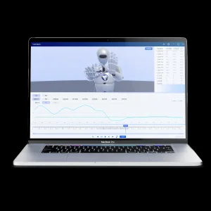
Best Valyze | Visualized Intelligent Motion Data Analysis Software for ce, Sports, Rehabilitation & Education Planning, Solution
Explore Solution →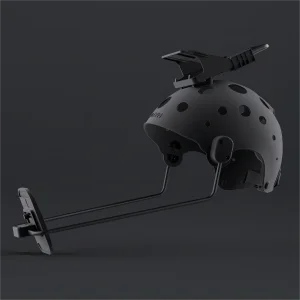
Best Virdyn VDFace Pro Capture System for Real Time Facial Capture with Face Capture Helmet Planning, Solution
Explore Solution →
Best Customize Your 3D Virtual Avatar - Personalized High-Quality and Fast Delivery | 3D Custom Avatar Services Planning, Solution
Explore Solution →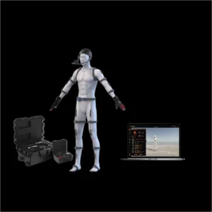
Design DreamsCap X1 Full-Body, Hand & Face Motion Capture for Humanoid Robot Data Collection & Teleoperation Planning, Solution
Explore Solution →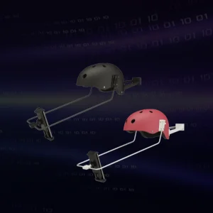
Best Virdyn VDFace Capture System for Real Time Facial Capture with Face Capture Helmet Planning, Solution
Explore Solution →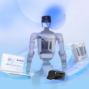
Design AI LingSi System | Robotic Intelligent Creation Platform & Intelligent Voice Interaction Planning, Solution
Explore Solution →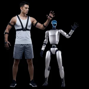
Best VDSuitMini Half-Body Mocap System | Robot Data Collection & Teleoperation | Free Software Planning, Solution
Explore Solution →Strategic Recommendations for Clinical Project Planning
Bridging biomechanical precision, artificial intelligence, and personalized medicine: A checklist for clinical leaders planning next-generation physical therapy infrastructure.
As healthcare systems transition toward value-based reimbursement frameworks, proving functional rehabilitation efficacy through continuous, objective metrics is paramount. Motion capture technology has transcended its original roots in cinematic entertainment, establishing itself as the empirical foundation of modern neuro-rehabilitation and orthopedic recovery tracking.
When selecting and designing a clinical motion analysis solution, hospital procurement boards, university researchers, and rehabilitation entrepreneurs should prioritize the following strategic pillars:
Clinical Accuracy & Repeatability
Insist on audited third-party validation studies against gold-standard optical stereophotogrammetry (Vicon/Qualisys) proving angular error below 1.0 degree across extended dynamic movements.
Patient Usability & Ergonomics
Prioritize rapid, non-invasive mounting protocols. Garments and harnesses must withstand continuous clinical wash cycles, accommodate amputations, and remain comfortable for elderly populations.
Interoperability & Open APIs
Ensure native export and streaming capabilities (BVH, FBX, CSV, UDP/TCP raw quaternion feeds) with direct support for MATLAB, Python, OpenSim, Unity, and HL7 healthcare standards.
Regulatory Readiness & Security
Verify that prospective hardware architectures possess pathways toward MDR/FDA compliance, with comprehensive encryption safeguarding delicate biographical and kinematic patient files.